In anesthetic practice, preoperative airway assessment seeks to identify patients at risk for difficult ventilation or intubation. The expansion of point-of-care ultrasound in recent years has increased interest in ultrasound airway evaluation before anesthesia and surgery, offering real-time, noninvasive visualization of airway anatomy and improving perioperative decision-making.
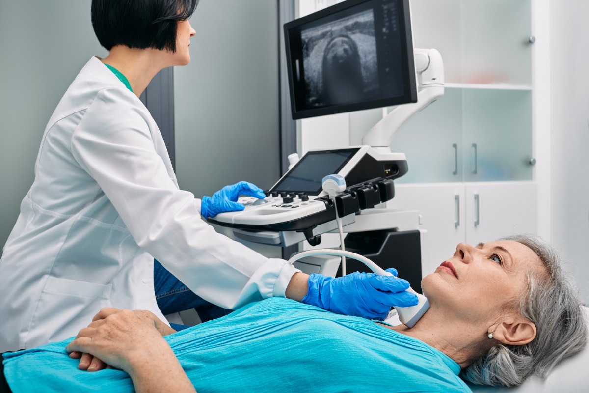
Ultrasound enables detailed assessment of upper airway structures, including the tongue, epiglottis, vocal cords, and trachea. It can also be used to measure anterior neck soft tissue thickness, which has been associated with difficult laryngoscopy. Studies have demonstrated that increased pre-tracheal soft tissue thickness at the level of the vocal cords correlates with higher grades of laryngoscopic difficulty. Additionally, ultrasound can identify anatomical variations, masses, or airway pathology that may not be apparent on physical examination.
One of the most widely studied applications of ultrasound for airway evaluation is the prediction of difficult intubation, which is a key clinical challenge for anesthesia professionals. Measurements such as the distance from the skin to the epiglottis, hyomental distance ratio, and tongue thickness have shown promise as objective predictors. While no single parameter has proven definitive, combining multiple ultrasound measurements with clinical assessment enhances predictive accuracy. This multimodal approach aligns with current trends in perioperative risk stratification.
Ultrasound is also valuable in identifying the cricothyroid membrane, particularly in patients with difficult anatomy such as obesity, neck deformities, or prior surgery. Accurate localization of this structure is critical in emergency airway situations requiring cricothyrotomy. Compared to palpation alone, ultrasound-guided identification has been shown to be more reliable and reproducible, reducing the risk of complications during front-of-neck access.
Another important application is confirmation of endotracheal tube placement. Ultrasound can rapidly detect tracheal versus esophageal intubation by visualizing tube passage and assessing lung sliding bilaterally. This technique is especially useful in situations where capnography may be unreliable, such as during cardiac arrest or low-perfusion states. Furthermore, ultrasound can aid in determining appropriate endotracheal tube size, particularly in pediatric populations, by measuring subglottic airway diameter.
However, clinicians should be aware of the limitations of using ultrasound for airway evaluation. Operator dependency remains a significant challenge, as accurate image acquisition and interpretation require training and experience. Additionally, variability in measurement techniques and lack of standardized protocols limit widespread adoption. Time constraints in emergency settings may also restrict its routine use, although rapid scanning protocols are being developed.
Recent literature supports the integration of airway ultrasound into patient evaluation before anesthesia and surgery, particularly in high-risk patients. It is increasingly incorporated into anesthesiology training programs and clinical guidelines as an adjunct rather than a replacement for traditional methods. Future research is focused on standardizing measurement techniques, validating predictive models, and improving accessibility through portable ultrasound devices.
Ultrasound is a useful tool for airway evaluation before anesthesia, enhancing the ability to predict difficult airways, guide interventions, and improve patient safety. When combined with conventional assessment methods, it provides a more comprehensive and objective evaluation. As technology advances and training becomes more widespread, ultrasound is likely to play an increasingly central role in perioperative airway management.
References
1. Kristensen MS, Teoh WH, Graumann O, Laursen CB. Ultrasonography for clinical decision-making and intervention in airway management: from the mouth to the lungs and pleurae. Insights Imaging. 2014;5(2):253-279. doi: 10.1007/s13244-014-0309-5.
2. Fulkerson JS, Moore HM, Anderson TS, Lowe RF. Ultrasonography in the preoperative difficult airway assessment. J Clin Anesth. 2017;37:52-59. doi: 10.1007/s10877-016-9888-7.
3. Adhikari S, Zeger W, Schmier C, et al. Pilot study to determine the utility of point-of-care ultrasound in the assessment of difficult laryngoscopy. Acad Emerg Med. 2011;18(7):754-758. doi: 10.1111/j.1553-2712.2011.01099.x.
4. Siddiqui N, Arzola C, Iqbal M, et al. Ultrasound is superior to landmark technique for identification of the cricothyroid membrane in subjects with poorly defined neck anatomy. Anesthesiology. 2018;129(6):1130-1139. doi: 10.1097/ALN.0000000000002454.
5. Gottlieb M, Holladay D, Peksa GD. Point-of-care ultrasound for the confirmation of endotracheal tube placement: a systematic review and meta-analysis. Ann Emerg Med. 2018;72(6):627-636. doi: 10.1016/j.annemergmed.2018.06.024.